
Aug 18, 2015
In our previous Newsletters, we have covered background and applications about Whole Slide Imaging (WSI). This post is a quick reminder of what WSI is, and here are 3 quick facts about WSI:
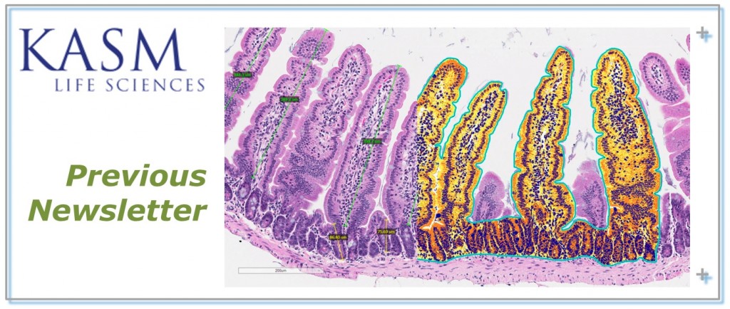
WSI converts histologically or immunohistochemically (IHC) stained glass slides into digital slides, as known as whole slide images (WSIs). One could perform similar tasks as she/he would with a light microscope on computer monitors. A researcher could zoom in or out of a region of interest in the office or with a laptop on the run. There is no more spending extra energy and time at searching a slide for re-capturing due to focusing issues and no more zipping images, as every corner of a tissue section is captured with crisp and sharpness on WSIs. One could take snapshots with desired magnification with embedded scale bars, and this saves time from scale calibration from images taken with conventional microscopes.
WSI opens doors of opportunity to study your experiments. Repeatable image analyses could be performed by computer algorithms in a timely manner for studying IHC slides. For analyzing histology or pathology slides, quantification such as length and area could be measured accurately on computer monitors. Furthermore, annotations on the WSIs could also be shared digitally to enhance education or conference call quality.
Want to learn more about WSI? Please fill the following form and we will send you a copy of our previous newsletters. Feel free to contact us for additional information!
July 23, 2015
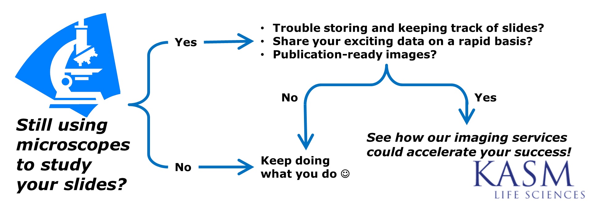
Do you constantly find unfocused and blurry tissue area from whole slide images from your in-house facility or high-throughput scanning providers? Our dedicated staff perform stringent QA/QC when preparing your Whole Slide Images. We ensure all tissue regions are captured with crisp and sharpness. Our dedication and expertise offer you the best Digital Pathology experience! How could we help to translate your pixels into meaningful solutions?
We provide imaging and consulting services (Custom Whole Slide Imaging, Confocal Microscopy and Image Analysis) to the Life Science sectors. We combine our extensive industrial and academic experiences to serve a diverse client base including academic laboratories, biotechnology, pharmaceutical, start-up bio/pharmaceutical, device and diagnostic companies. KASM solutions include market research, technology assessment, project planning and management, CRO/CMO sourcing and technology transfer along with scientific writing and presentation.We effectively communicate our findings to all levels of experience ranging from the technologist to the CEO. We provide scientific, technical and business advice with a keen appreciation for critical business and organizational considerations ensuring that projects are delivered with professionalism on time and within budget. In addition to the principals, we utilize a network of consultants based on project requirements.
Stay tune for more industry and KASM news!