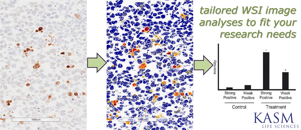
We offer a number of tested and refined Image Analyses to extract meaningful information out of whole slide images (WSIs). In addition to studying expression patterns from IHC slides, customizable algorithms reveal precise and quantitative data that is accurate and repeatable. WSI Image analysis helps to address, but not limited to the following: Where and how much staining is there? Where and how many objects are there in tumor cells? How much staining are there in nuclei and membranes?
WSI also enables accurate and high-throughput quantification for histological slides. For example, Scar Elevation Index could be efficiently measured from rabbit wound healing models.
To learn more about WSI Image Analysis, check out our Blog posts or Contact us for more information!